Stroke is more commonly associated with older adults. However, there is a rare condition that can cause stroke in both children and young adults, known as moyamoya disease. This disease causes narrowing of the major blood vessels in the brain, reducing blood flow to brain tissue. In its early stages, the symptoms often go unnoticed, so the condition is frequently diagnosed only after a stroke or other neurological complications have occurred.
Although considered rare, moyamoya disease should be recognized as early as possible because it can significantly increase the risk of stroke, impaired brain function, and a reduced quality of life if left untreated.
Moyamoya disease is a cerebrovascular disorder characterized by progressive narrowing of the internal carotid arteries and their major branches at the base of the brain. This narrowing reduces blood flow to the brain, preventing brain tissue from receiving adequate oxygen and nutrients.
In response, the body forms a network of tiny blood vessels as alternative pathways (collateral vessels) to maintain blood flow to the brain. However, these collateral vessels are small and fragile, making them unable to fully replace the function of the main arteries. They are also prone to rupture, which can lead to bleeding in the brain.
On Digital Subtraction Angiography (DSA), this network of tiny blood vessels appears as a cloud of smoke. In Japanese, the word "moyamoya" means "a puff of smoke," which is how the disease got its name.
Moyamoya disease may affect one side or both sides of the brain. However, in most patients, the narrowing gradually progresses to involve both sides over time if left untreated.
The symptoms of moyamoya disease vary from person to person, depending on age and the severity of blood vessel narrowing. Symptoms develop either because of reduced blood flow to the brain or due to rupture of the fragile collateral blood vessels.
In children, the disease most commonly presents as a Transient Ischemic Attack (TIA) or an ischemic stroke. In adults, intracranial hemorrhage (hemorrhagic stroke) is also commonly observed in addition to ischemic stroke.
Symptoms that should not be ignored include:
In some patients, symptoms may be triggered by strenuous physical activity, crying, fever, coughing, or hyperventilation, as these conditions can further reduce blood flow to the brain.
To date, the exact cause of moyamoya disease remains unknown. However, several factors are known to be associated with an increased risk of developing the condition.
Genetic factors play an important role in the development of moyamoya disease. One of the genes most commonly associated with the disease is RNF213, particularly among East Asian populations. However, not everyone who carries this genetic variant will develop moyamoya disease.
The risk is also higher in individuals who have a family member—especially a parent or sibling—with moyamoya disease.
Moyamoya disease most commonly occurs in two age groups: children between 5 and 10 years old and adults between 35 and 50 years old. In children, the disease typically presents as an ischemic stroke or TIA. In adults, symptoms may include ischemic stroke, intracranial hemorrhage, or chronic headaches.
Several genetic disorders are known to increase the risk of developing moyamoya disease, including:
These conditions may affect the development and structure of blood vessels, increasing susceptibility to narrowing of the brain's arteries.
In addition to moyamoya disease, there is another condition known as moyamoya syndrome, which refers to blood vessel narrowing that resembles moyamoya disease but occurs secondary to another underlying medical condition.
Several conditions associated with moyamoya syndrome include:
The role of environmental factors is still being investigated. To date, no environmental factor has been conclusively proven to cause moyamoya disease. However, it is believed that an interaction between genetic and environmental factors may contribute to the development of the disease.
The diagnosis of moyamoya disease is established through a combination of medical history taking, physical examination, neurological examination, and brain imaging studies.
Diagnostic tests that may be performed include:
An accurate diagnosis is essential so that appropriate treatment can be initiated before recurrent strokes occur.
The goals of treatment are to reduce the risk of stroke, preserve brain function, and improve the patient's quality of life.
Depending on the patient's condition, doctors may prescribe medications such as:
It is important to note that medications cannot reverse the narrowing of the blood vessels. Instead, they help reduce the risk of complications.
Surgery is the primary treatment for most patients with moyamoya disease. The purpose of this procedure is to create a new pathway to improve blood flow to the brain. Common surgical techniques include:
In some patients, both techniques may be combined to achieve the best possible outcome. The choice of surgical procedure depends on the patient's age, the condition of the blood vessels, imaging findings, and the neurosurgeon's clinical judgment.
Numerous studies have shown that revascularization surgery can reduce the risk of recurrent stroke while improving patients' long-term quality of life.
Moyamoya disease is a cerebrovascular disorder that can cause stroke in both children and adults. Although it is considered a rare condition, the disease is progressive, making early diagnosis and timely treatment essential to prevent recurrent strokes, permanent brain damage, and a decline in quality of life.
Do not ignore symptoms such as sudden weakness on one side of the body, speech difficulties, seizures, or recurrent headaches, especially when they occur in children or young adults. The earlier moyamoya disease is recognized, the greater the opportunity to preserve brain function and prevent more serious complications.
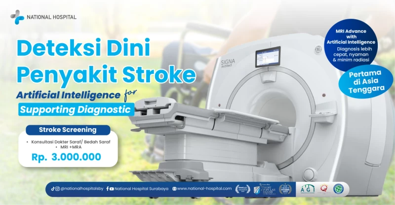
The National Hospital Neuroscience Center is supported by an experienced team of neurologists and neurosurgeons, along with advanced diagnostic facilities such as MRI, CT Scan, and Digital Subtraction Angiography (DSA) to help establish an accurate diagnosis and determine the most appropriate treatment plan for each patient.






 info@national-hospital.com
 Suggestion
 Login
Login.png)