Magnetic Resonance Imaging (MRI) is one of the most important medical imaging methods that helps doctors diagnose a wide range of health conditions. Unlike some other imaging examinations, MRI uses a powerful magnetic field and radio waves to produce highly detailed images of organs and body tissues without exposing patients to radiation.
As technology continues to advance, MRI systems have become increasingly sophisticated. One of the latest innovations is the 3 Tesla (3T) MRI equipped with Artificial Intelligence (AI), which delivers higher-quality images while significantly improving examination efficiency.
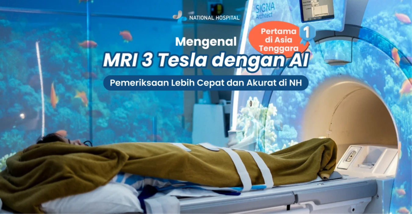
National Hospital is the first hospital in East Java to introduce 3 Tesla MRI with AI technology, supporting more accurate and comfortable diagnostic services for patients. But what exactly is MRI, and who should undergo an MRI examination? Read on for the complete explanation!
MRI is a radiological examination used to visualize the structure of organs, soft tissues, blood vessels, and the nervous system in great detail. This examination is often recommended when doctors require information that cannot be adequately obtained through other imaging methods such as X-rays or ultrasound.
MRI machines are large enough to accommodate the patient's entire body. The tube-shaped magnet inside the MRI machine generates a strong magnetic field that realigns the protons found in water molecules within the body's tissues. The radio waves emitted by the machine then help create three-dimensional (3D) images.
These images are displayed as cross-sectional slices, allowing doctors to examine each layer of body tissue in greater detail. Since MRI does not use ionizing radiation, it is considered a safe option, especially for patients who require repeated imaging examinations to monitor chronic health conditions.
Below are the stages of the MRI process in more detail:
The human body is largely composed of water (H₂O), meaning it contains billions of hydrogen atoms. An MRI machine resembles a large tube equipped with a giant magnet. While lying inside the MRI scanner, the powerful magnetic field forces the protons (the nucleus of hydrogen atoms) in the body to align in a specific direction.
The MRI machine emits short bursts of radio waves toward the area being examined. These radio waves disrupt the alignment of the protons and cause them to shift. Once the radio waves are turned off, the protons return to their original aligned position while emitting energy signals.
When the radio waves stop, the protons return to their aligned position while releasing energy in the form of radio signals. These signals are captured by sensors inside the MRI machine and transmitted to a computer. Different tissues such as fat, muscle, blood, and fluid emit signals with distinct characteristics.
The receiving coils inside the MRI scanner detect these frequencies and send them to a computer. The computer then converts the signal data into highly detailed three-dimensional (3D) grayscale images, ranging from deep black through multiple shades of gray to bright white. This is what enables MRI to provide extremely detailed anatomical images.
Conventional MRI (1.5 Tesla) produces excellent image quality that is suitable for routine diagnostic purposes. Meanwhile, AI 3 Tesla MRI delivers sharper, higher-resolution (HD/ultra-detailed) images, making it ideal for detecting very small lesions or abnormalities and providing better visualization of anatomy.
Compared to the longer examination time required for a 1.5 Tesla MRI, the AI 3 Tesla MRI performs scans up to 30% faster. This allows patients, especially those with claustrophobia or fear of confined spaces, to undergo a more comfortable examination due to the shorter scanning time.
With this MRI technology, patient comfort is enhanced without compromising diagnostic precision, accuracy, or early disease detection, enabling doctors to provide the most appropriate treatment.
MRI can help evaluate various health conditions. Doctors usually recommend an MRI for patients experiencing conditions such as:
MRI is intended to examine specific organs of the body. Not every organ can be evaluated using MRI. Commonly examined organs include:
MRI is used to diagnose several conditions affecting the brain and spinal cord, including brain aneurysms, brain injuries, eye or ear disorders, pinched nerves, multiple sclerosis (MS), stroke, and brain or spinal tumors.
MRI is used in patients with heart-related complaints to evaluate several conditions, including the anatomical structure of the heart, blockages or abnormalities in blood vessels, blood flow through arteries and veins, congenital heart disease, damage from heart attacks, heart function, infections, tumors, and inflammation.
MRI is frequently used to detect various bone and joint disorders, including bone cancer, bone infections, and joint injuries. MRI utilizes radio waves and highly sensitive magnetic technology to provide detailed and accurate images of bone and joint structures.
MRI is also recommended for patients at high risk of developing breast cancer. In addition to mammography, MRI is often used as a complementary examination to detect cancer cells at an earlier stage. MRI can also be performed to evaluate prostate disorders. Other internal organs commonly examined with MRI include the liver, spleen, pancreas, kidneys, ovaries, uterus, and testes.
As part of its commitment to providing innovative diagnostic services, National Hospital has introduced East Java's first 3 Tesla (3T) MRI equipped with Artificial Intelligence (AI). Launched in 2023 through a collaboration with GE HealthCare, this technology is designed to deliver faster, more accurate, and more comfortable imaging examinations for patients.
The 3 Tesla MRI produces higher-resolution images than conventional MRI systems. Supported by AI-powered algorithms, the scanning process can be completed up to approximately 60% faster while simultaneously producing sharper and clearer images. This enables doctors to detect diseases earlier, establish more accurate diagnoses, and monitor patients' responses to treatment more effectively.
This technology is particularly valuable for various diagnostic needs, especially conditions requiring immediate medical attention, such as stroke. With superior image quality and shorter examination times, doctors can obtain critical diagnostic information more quickly, supporting timely and appropriate medical decision-making.
Supported by experienced radiologists, professional radiographers, and modern facilities, the 3 Tesla MRI with AI service at National Hospital reflects the hospital's commitment to delivering high-standard diagnostic services that help patients receive prompt, accurate, and comprehensive medical care.






 info@national-hospital.com
 Suggestion
 Login
Login