Breast cancer is one of the health problems that requires serious attention. One important step to increase the chances of successful treatment is to detect abnormalities in the breast as early as possible, even before symptoms appear. Currently, breast examinations do not rely on just one method.
In addition to clinical examination and mammography, other imaging technologies are available that can be used as supplementary examinations according to the patient’s condition and needs. One of them is Invenia ABUS (Automated Breast Ultrasound System). Here is a complete explanation!
ABUS stands for Automated Breast Ultrasound System, an automated breast ultrasound technology that uses a scanning system to produce three-dimensional (3D) images of breast tissue. This technology is designed as a supplementary examination for breast cancer screening, particularly for women with dense breast tissue.
Unlike conventional breast ultrasound, which uses a transducer operated by an examiner, ABUS uses an automated scanning system to produce breast images in a more standardized and consistent manner. The examination results consist of a collection of 3D images that can then be evaluated by a radiologist.
Invenia ABUS, manufactured by GE HealthCare, is one of the Automated Breast Ultrasound technologies developed as a supplementary examination for women with dense breasts. The FDA lists Invenia ABUS as an adjunct examination to mammography for breast cancer screening in certain populations with dense breast tissue.
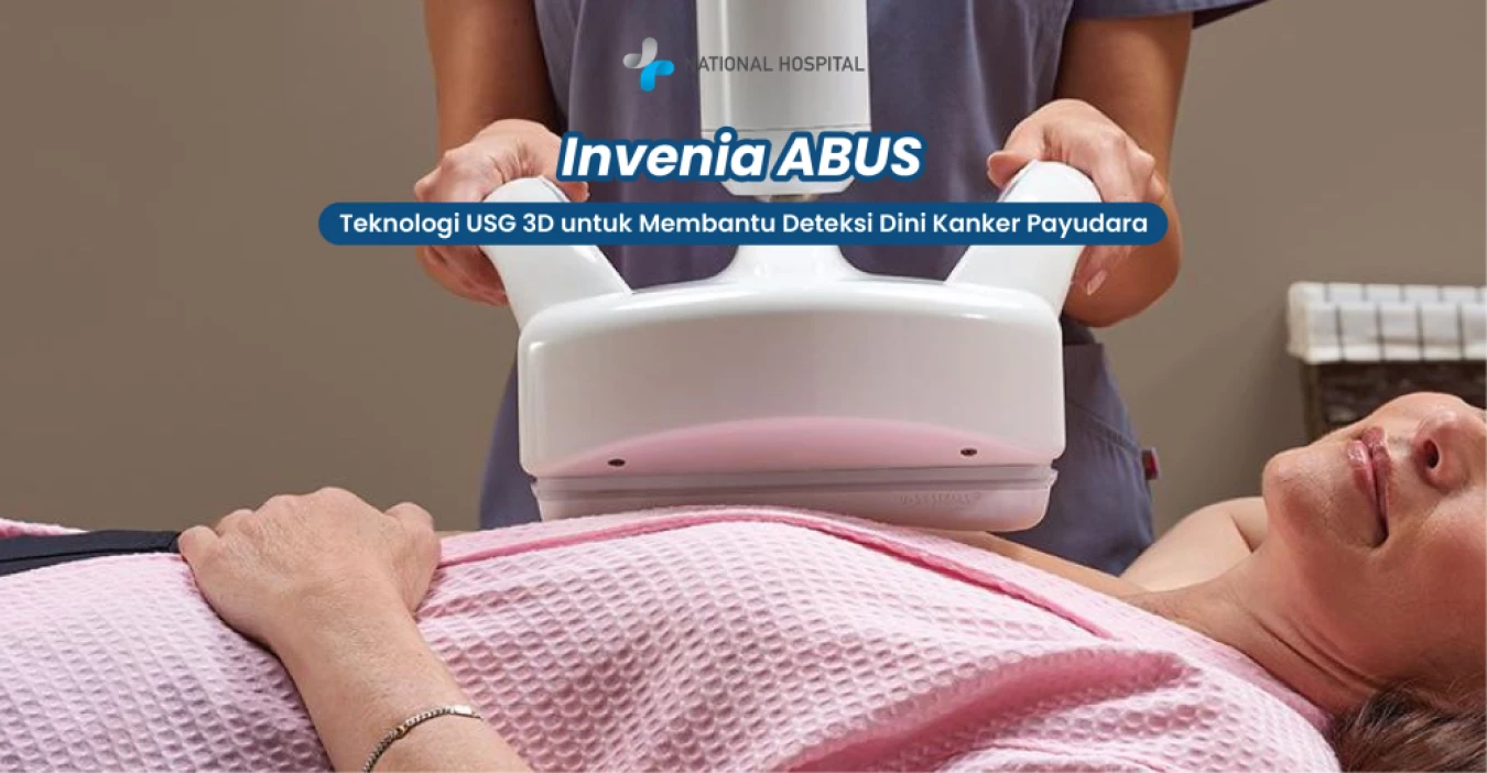
Invenia ABUS is a non-invasive procedure that does not use ionizing radiation. The patient will be asked to lie down, after which the breast will be positioned for scanning using a specialized transducer. The ABUS system will automatically perform the scan to produce a collection of 3D images of the breast tissue.
Because the image acquisition process is performed automatically and in a standardized manner, the examination can produce consistent images that can then be reviewed and analyzed by a radiologist. The image acquisition process for Invenia ABUS 2.0 can be completed in approximately 15 minutes. However, the overall examination duration may vary depending on the patient’s condition and the healthcare facility’s service flow.
Invenia ABUS technology is also equipped with artificial intelligence (AI)-based features that can assist in the image acquisition and interpretation process. Nevertheless, AI functions as a supporting tool and does not replace the physician. Examination results still need to be evaluated by a radiologist, taking into consideration the patient’s clinical condition and overall examination results.
Some characteristics of Invenia ABUS that make this technology an interesting option for supplementary examination include:
In clinical data cited by GE HealthCare, the use of Invenia ABUS as a supplementary examination in women with dense breasts showed a 35.7% increase in cancer detection compared with mammography alone. This figure comes from research conducted in a specific population and examination setting, and therefore should not be interpreted as meaning that ABUS will increase detection by the same amount in every individual.
Each breast examination method has its own advantages and limitations. One factor that needs to be considered is breast tissue density. Breasts are composed of fatty tissue, glandular tissue, and connective tissue. On mammography, dense breast tissue appears white. Some abnormalities, including breast cancer, may also appear white, which under certain conditions can make them more difficult to identify among dense breast tissue.
Therefore, for women with dense breasts, doctors may consider supplementary imaging examinations based on each patient’s condition and risk factors. One of them is Invenia ABUS (Automated Breast Ultrasound System), which uses automated ultrasound technology to produce 3D images of breast tissue.
It is important to understand that Invenia ABUS does not replace mammography. ABUS was developed as a supplementary screening examination for mammography in certain patient groups, particularly women with dense breast tissue. Therefore, mammography continues to play an important role in breast cancer screening, while ABUS may provide additional information about breast tissue.
The choice of examination should be tailored to each patient’s condition, including age, breast density, risk factors, medical history, presence of symptoms, and the doctor’s recommendation. With the right approach, various examination methods can complement one another to support early detection of breast cancer.
Invenia ABUS may primarily be considered as a supplementary examination for women with dense breasts, based on the doctor’s assessment. However, this does not mean that every woman with dense breasts must undergo ABUS. The decision to undergo the examination should take the patient’s overall condition into consideration. Doctors may consider several factors, such as:
For women who have a lump, changes in breast shape, skin changes, nipple discharge, or other suspicious symptoms, diagnostic examinations should be performed based on the doctor’s evaluation. ABUS for screening is not intended to replace medical evaluation in patients who have symptoms.
Invenia ABUS (Automated Breast Ultrasound System) is an automated 3D breast ultrasound technology that can be used as a supplementary examination, particularly for women with dense breast tissue.
This technology produces standardized images of breast tissue and does not use ionizing radiation. However, Invenia ABUS is not a replacement for mammography. The need for ABUS examination should be determined based on the patient’s condition, breast density, risk factors, medical history, and the doctor’s recommendation.
As part of its commitment to providing healthcare services supported by modern technology, National Hospital provides Invenia ABUS examination services to support breast cancer screening and early detection, particularly for patients who require supplementary examinations based on medical indications.
The good news is that a Medical Check-Up promotion with a free consultation with an oncology specialist at National Hospital is currently available. Take advantage of this opportunity to learn more about your breast health and find out whether ABUS examination is appropriate for your needs.






 info@national-hospital.com
 Suggestion
 Login
Login