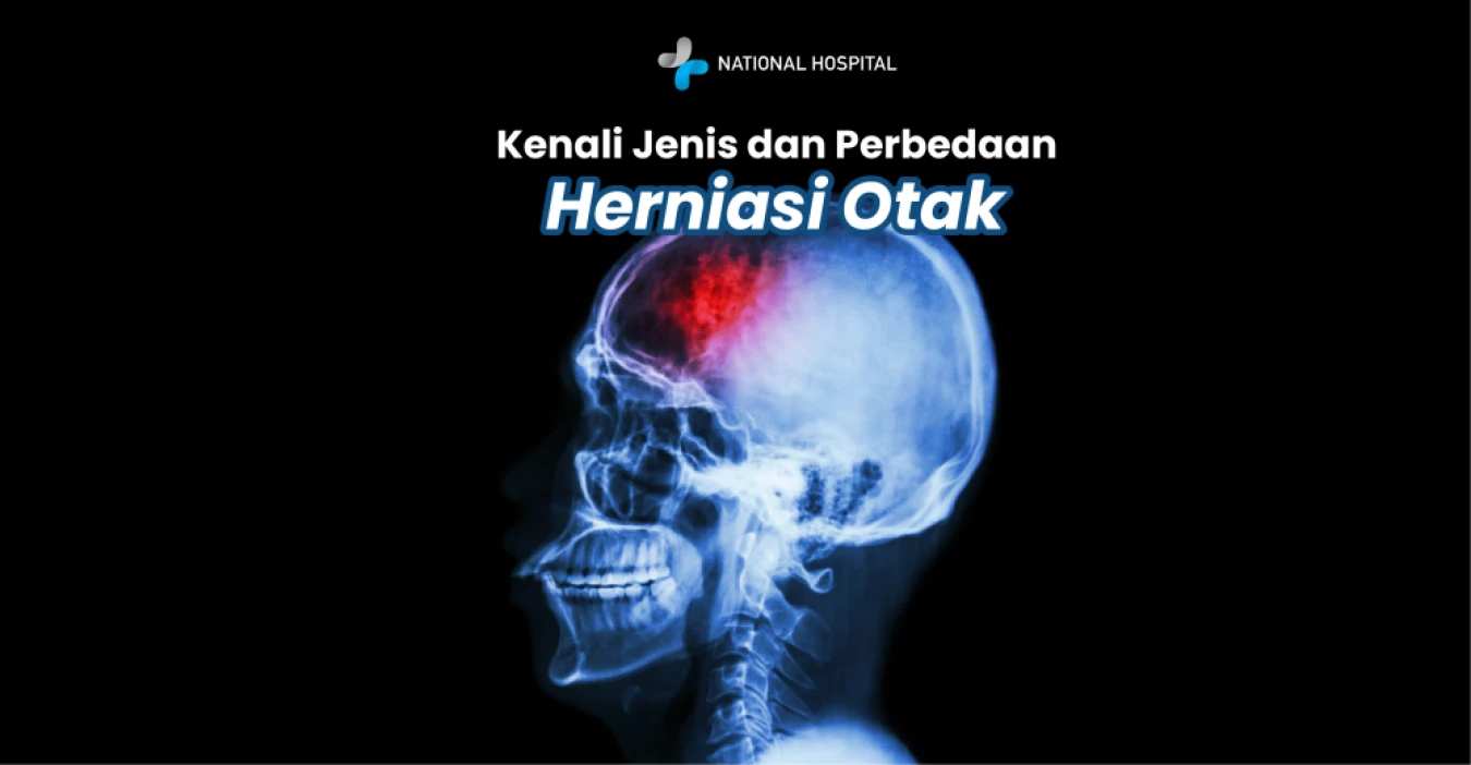
Have you ever heard the term “brain shift”? In the medical world, this dangerous condition is known as brain herniation. It occurs when brain tissue is compressed and displaced from its normal position within the skull.
This displacement is generally caused by swelling, tumors, or bleeding in the head that drastically increases pressure inside the skull (elevated intracranial pressure).
This condition is extremely serious and life-threatening because the pressure can compress vital parts of the brain, such as the brainstem, which controls breathing and heart rate.
Given the high risk, it is very important to understand the different types of brain herniation and their differences. With better understanding, faster diagnosis and treatment can be carried out.
Brain herniation is classified into several types based on the location of brain tissue displacement. Below are the five main types you should know:
This is the most common type of brain herniation. It occurs when brain tissue shifts downward beneath the tough membrane in the midline of the brain called the falx cerebri.
Brain tissue shifts downward beneath the falx cerebri.
Often causes midline shift of the brain, which can be seen on CT scan as deviation of the septum pellucidum, and may trigger weakness on the opposite side of the body.
Depends on the degree of displacement. A shift greater than 15 mm is often associated with poor outcomes.
This type involves the temporal lobe of the brain and can occur in two forms: descending and ascending.
Descending transtentorial (uncal): Part of the temporal lobe (the uncus) shifts downward, compressing the oculomotor nerve (cranial nerve III) and the brainstem.
Ascending transtentorial: The cerebellum and brainstem shift upward.
Typical signs include a dilated and fixed pupil (non-reactive to light) on the same side as the herniation, drooping eyelid (ptosis), and abnormal eye position. Severe pressure can cause weakness on the same side of the body and decreased level of consciousness.
This type occurs when the central part of the brain (the diencephalon and temporal lobes) shifts downward.
The central brain tissue moves downward through the tentorial notch.
Initially, pupils may be small and reactive, but they can rapidly become dilated. This condition compresses the brainstem, which can lead to breathing disturbances, abnormal motor responses, and death if not treated immediately.
This is often referred to as “coning.”
The lowest part of the cerebellum (the cerebellar tonsils) shifts downward through the large opening at the base of the skull (foramen magnum).
Compresses the brainstem, which controls vital functions such as breathing and heart rate. Symptoms include decreased consciousness, irregular breathing patterns, dilated pupils, and a high risk of cardiac arrest.
This type differs from others because the brain tissue shifts outward.
The brain protrudes outward through a surgical opening or an open skull defect due to extremely high intracranial pressure.
Usually associated with severe head trauma and open injuries. Symptoms vary widely and often require emergency surgery.
Brain herniation is a medical emergency that occurs when brain tissue is forced beyond its normal boundaries due to increased intracranial pressure. This condition can cause severe brain damage or death if not treated promptly. Therefore, early detection is crucial, and Brain MRI is one of the most accurate examinations to detect brain herniation.
With Brain MRI screening, doctors can visualize brain tissue displacement, brainstem compression, or the presence of masses that trigger herniation. Early detection allows for immediate medical or surgical intervention to prevent serious complications and improve recovery outcomes.
National Hospital Surabaya provides comprehensive and modern Brain MRI services, supported by experienced medical professionals, to ensure accurate diagnosis and appropriate management for patients at risk of brain herniation. Routine screening or immediate examination when symptoms appear is strongly recommended to maintain brain health and prevent emergencies.
Brain herniation is a critical emergency condition that requires immediate medical attention. Common symptoms of increased intracranial pressure that can trigger herniation include severe headache, intense nausea and vomiting, and decreased consciousness. If you or a family member experiences these symptoms, do not delay seeking medical help.
Because it is a life-threatening condition that requires rapid and precise diagnosis and treatment, brain herniation must be evaluated by specialists. We strongly recommend consulting a neurosurgeon for an accurate diagnosis and appropriate medical management, which may include surgical intervention to relieve pressure on the brain.
Neurosurgeons have the expertise and experience to manage complex brain and spinal conditions. Prompt and appropriate treatment—especially in critical conditions such as brain herniation—greatly influences outcomes and recovery chances.
If you are in the Surabaya area and require rapid neurosurgical care, you may consider seeking further information about the services and facilities available at National Hospital. Always prioritize emergency medical care if you experience signs of brain herniation.






 info@national-hospital.com
 Suggestion
 Login
Login