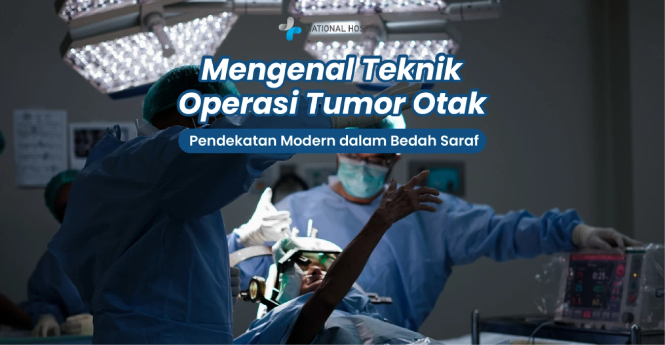
Brain tumor surgery is one of the most complex neurosurgical procedures, aiming to safely remove brain tumors while preserving the patient’s neurological functions. This procedure is performed to reduce pressure on the brain, manage symptoms caused by tumors such as headaches and seizures, obtain an accurate diagnosis, and prevent further disease progression.
With advances in medical technology, various surgical techniques are now available to treat brain tumors with greater safety, precision, and minimal risk to critical brain functions. The selection of surgical techniques is tailored to each patient based on the tumor’s location, size, type, overall health condition, and potential risks to important neurological functions. With the right approach, tumor removal can be performed optimally while maintaining the patient’s quality of life.
The following are several brain tumor surgery techniques commonly used in modern neurosurgical practice.
Craniotomy is one of the most commonly performed procedures for brain tumor surgery. This procedure involves removing a portion of the skull (cranium) to provide direct access to the brain and the tumor that needs to be removed. The primary goal of a craniotomy is to allow neurosurgeons to remove as much of the tumor as possible while preserving the patient’s neurological function.
In addition to tumor removal, craniotomy can also help relieve increased intracranial pressure caused by the tumor mass and alleviate various neurological symptoms such as headaches, seizures, vision problems, and limb weakness.
Although effective, this procedure carries potential risks, including bleeding, infection, postoperative seizures, temporary or permanent neurological deficits, and brain swelling.
Endoscopic Endonasal Transsphenoidal Surgery (EETS) is a minimally invasive surgical technique used to remove tumors located at the skull base and pituitary gland region through the nasal cavity without opening the skull.
This technique is commonly used to treat pituitary adenomas, craniopharyngiomas, chordomas, and certain types of skull base meningiomas that can be accessed through the endonasal approach.
The main advantage of this technique is that it does not require an incision on the scalp, resulting in no external surgical scar. In addition, patients generally experience less postoperative pain, shorter hospital stays, and faster recovery compared to conventional craniotomy.
However, this procedure still carries potential risks, including cerebrospinal fluid leakage, hormonal disorders, infection, and visual impairment in certain cases.
Skull Base Surgery refers to a group of surgical techniques used to treat tumors located at the skull base, an anatomically complex area due to its close proximity to the brain, cranial nerves, major blood vessels, and other vital structures.
Skull base surgery may be performed through an open skull base approach, an endoscopic endonasal approach, or a combination of both techniques involving multidisciplinary collaboration.
This approach allows surgeons to access tumors located in difficult-to-reach areas while minimizing injury to normal brain tissue. Due to its high complexity, skull base surgery requires careful planning and support from advanced imaging technology and modern surgical navigation systems.
Keyhole Brain Surgery is a minimally invasive neurosurgical technique that uses a small skull opening, generally around 2–3 centimeters, to access and remove brain tumors.
Instead of creating a large opening in the skull, surgeons utilize natural anatomical pathways to reach the tumor location through a less invasive approach.
This technique is commonly used for small to medium-sized brain tumors, selected skull base tumors, brain cysts, and other lesions that can be accessed through minimally invasive routes.
The advantages of this technique include reduced tissue trauma, less postoperative pain, lower risk of infection, and faster recovery. However, not all tumors are suitable for this approach, as the decision depends on the tumor’s size, location, and characteristics.
A brain tumor biopsy is a procedure performed to obtain a tissue sample from a tumor for microscopic examination in the laboratory. This examination aims to determine the type of tumor, assess its grade of malignancy, and assist in planning further treatment, including radiotherapy, chemotherapy, targeted therapy, or additional surgical procedures.
In some cases, a biopsy may be performed as a standalone procedure or combined with tumor removal surgery.
Common types of brain tumor biopsy include:
Biopsy offers the advantage of being a relatively minimally invasive procedure with high diagnostic accuracy. However, like other surgical procedures, it still carries risks such as bleeding, infection, and mild neurological complications in certain conditions.
Awake Craniotomy is a brain tumor surgery technique in which the patient remains conscious during certain stages of the procedure. This technique is primarily used for tumors located near areas of the brain responsible for essential functions such as speech, language, movement, and memory.
During surgery, the medical team performs brain mapping to identify critical functional areas that must be preserved. At this stage, patients may be asked to perform simple tasks such as speaking, counting, reading, or moving certain body parts, allowing the surgical team to monitor neurological function directly.
The advantages of this technique include reducing the risk of permanent speech impairment, preserving motor and cognitive functions, and enabling more optimal tumor removal with improved safety.
However, this procedure requires excellent cooperation between the patient, neurosurgeon, and an experienced anesthesia team.
In addition to various surgical techniques, modern neurosurgery is supported by Intraoperative Monitoring (IOM) technology, a system that provides real-time monitoring of nerve function during surgery.
This technology is not a surgical technique itself but serves as a supporting tool that can be applied during various neurosurgical procedures.
During its application, electrodes are placed on specific areas of the body to monitor nerve and muscle activity throughout surgery. The goal is to detect potential neurological impairment as early as possible, allowing the surgical team to take immediate corrective action if needed.
The use of IOM helps reduce the risk of postoperative paralysis, preserve sensory and motor functions, improve surgical safety in complex locations, and enable more complete tumor removal while maintaining the patient’s neurological function.
Every brain tumor patient has a unique condition, meaning there is no single surgical technique suitable for all cases. The choice of surgical method must consider various factors, including tumor location, size, type, relationship with critical brain structures, patient health condition, and treatment goals.
With the support of modern technology, careful surgical planning, and collaboration among experienced multidisciplinary teams, brain tumor treatment can now be performed with increasingly improved safety and success rates.
The primary goal of treatment remains the same: to remove the tumor as effectively as possible while preserving the patient’s quality of life and neurological function to the greatest extent possible.
If you or your family experience symptoms that may indicate a brain tumor, such as persistent headaches, seizures, vision problems, limb weakness, or other changes in neurological function, consult a neurosurgeon promptly for proper evaluation and treatment.
National Hospital, through the National Hospital Neuroscience Center (NHNC), provides comprehensive brain tumor management services supported by experienced specialist doctors, advanced diagnostic technology, and a range of the latest neurosurgical techniques. Trust your health and well-being with National Hospital.






 info@national-hospital.com
 Suggestion
 Login
Login